- VRML model of Purkinje cell (generated from .p file)
- VRML model of cerebellar cortex slice
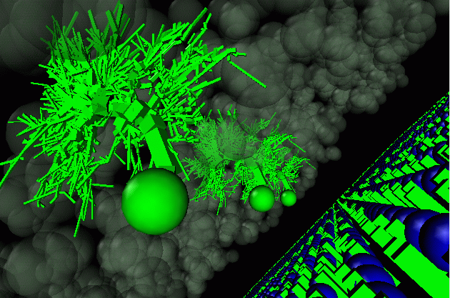
And another shot - the three green branching structures
are the Purkinje cells, with a cloud of inhibitory stellate cells around them. On the
right of the picture is the Golgi layer (blue spheres), with the mossy fiber input
also shown (green cuboids). This shot doesn't show the mass of parallel fibers.
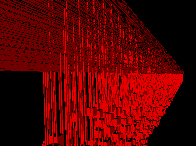
A picture of the slice of parallel fibers stored
by one processor. The granule cell somas are shown as red cubes, and their axons rise
to a T junction and extend in parallel for 2.5mm in each direction.
This shows about 3000 of the total of 240000 parallel fibers in
the simulation.
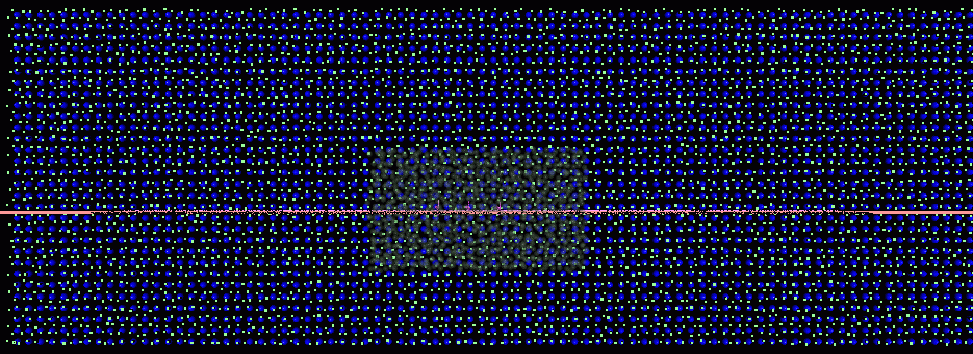
The top view of the simulated network. The grid of
blue Golgi cell somas, and green mossy fiber inputs is shown. In the centre is a
cloud of inhibitory Stellate cells, and just visible in the centre of this cloud
are three purple Purkinje cells. The red horizontal line crossing the picture is
one slice of parallel fibers.

Three slices of red granule cells are shown here, in this view along the line of the
parallel fibers (axons not shown). These are the leftmost, rightmost and
central slices. In the model, all the space between these three is filled
with granule cells at the same density as the slices (there are a total of 80
slices).
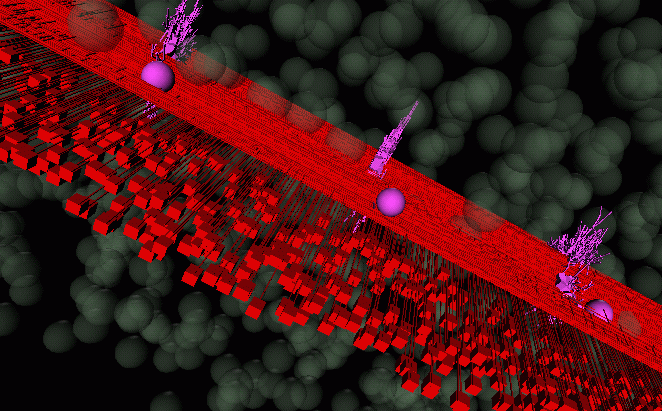
A shot from below, showing the (red) parallel fibers intersecting with the (purple)
Purkinje cells.